Aflutter Ecg. Atrial flutter is an abnormal heart rhythm that occurs in the atria of the heart. Atrial flutter is an abnormal cardiac rhythm caused by rapid atrial activity usually from reentry atrial circuits. Typical ecg findings include the presence of p waves and qrs complexes that have no association with each other , due to the atria and ventricles functioning independently. Electrocardiography (ecg or ekg) frequently makes the diagnosis by showing saw tooth flutter waves in several (ii, iii. For our 4th lesson, we continue our look at atrial and junctional arrhythmias.we start off talking about one of the most common arrhythmias that you will. Typical atrial flutter is widely known and recognised easily by clinical cardiologists however it does not always present a typical electrocardiographic pattern. How to make the difference between atrial fibrillation (afib) and atrial flutter and in particular between atypical atrial flutter and coarse atrial fibrillation. Atrial flutter causes characteristic ecg changes, as discussed below. Atrial flutter is a supraventricular tachycardic arrhythmia that tends to occur in individuals of an advanced age, although it is linked to endurance sports, also. Sawtooth baseline → flutter waves. The ecg criteria to diagnose atrial flutter are discussed including clockwise and counterclockwise, typical vs atypical atrial flutter, and different conduction patterns such as 1:1, 2:1, 3:1, 4:1 and 5:1. Afib and atypical aflutter requires more expertise and radiofrequency ablation has lower success rate. Differences between ecg wave strip patterns. Atrial flutter is diagnosed by you medical history, history of symptoms, and a physical exam. During atrial flutter the atria depolarize in an organized circular movement.
Aflutter Ecg , The Basics Of Atrial Flutter | Healing Heart Disease With Nurse Phyllis
Atrial flutter - Symptoms ,Pictures ,Causes And Treatment. Differences between ecg wave strip patterns. Atrial flutter is a supraventricular tachycardic arrhythmia that tends to occur in individuals of an advanced age, although it is linked to endurance sports, also. Electrocardiography (ecg or ekg) frequently makes the diagnosis by showing saw tooth flutter waves in several (ii, iii. The ecg criteria to diagnose atrial flutter are discussed including clockwise and counterclockwise, typical vs atypical atrial flutter, and different conduction patterns such as 1:1, 2:1, 3:1, 4:1 and 5:1. Atrial flutter is an abnormal heart rhythm that occurs in the atria of the heart. Sawtooth baseline → flutter waves. How to make the difference between atrial fibrillation (afib) and atrial flutter and in particular between atypical atrial flutter and coarse atrial fibrillation. Typical atrial flutter is widely known and recognised easily by clinical cardiologists however it does not always present a typical electrocardiographic pattern. Atrial flutter causes characteristic ecg changes, as discussed below. Typical ecg findings include the presence of p waves and qrs complexes that have no association with each other , due to the atria and ventricles functioning independently. Afib and atypical aflutter requires more expertise and radiofrequency ablation has lower success rate. Atrial flutter is an abnormal cardiac rhythm caused by rapid atrial activity usually from reentry atrial circuits. During atrial flutter the atria depolarize in an organized circular movement. Atrial flutter is diagnosed by you medical history, history of symptoms, and a physical exam. For our 4th lesson, we continue our look at atrial and junctional arrhythmias.we start off talking about one of the most common arrhythmias that you will.
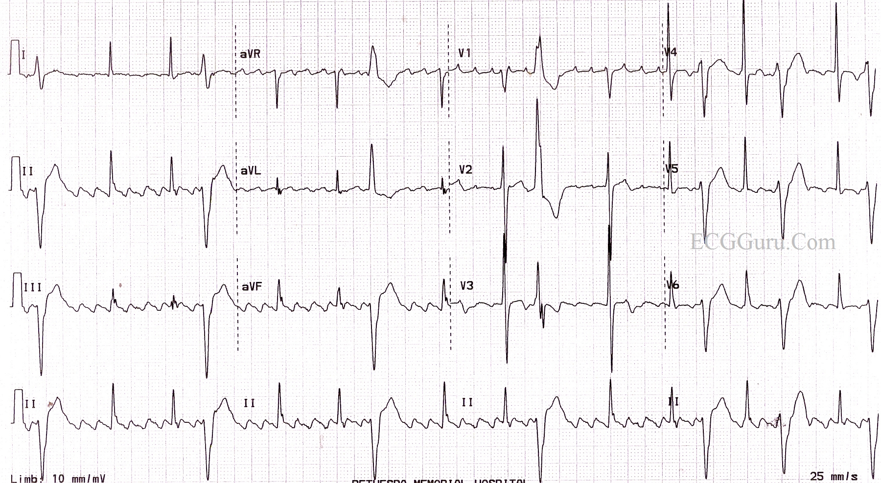
This ecg provides an example of atrial flutter with variable conduction.
Electrocardiography (ecg or ekg) frequently makes the diagnosis by showing saw tooth flutter waves in several (ii, iii. Atrial flutter is the second most common pathological tachyarrhythmia. Typical atrial flutter is widely known and recognised easily by clinical cardiologists however it does not always present a typical electrocardiographic pattern. 'coarse afib' has an f wave amplitude> 0.5 mm, which can mimic. Electrocardiography (ecg or ekg) frequently makes the diagnosis by showing saw tooth flutter waves in several (ii, iii. Atrial flutter causes characteristic ecg changes, as discussed below. Treatment of atrial flutter includes defibrillation of the heart and medication. It may be difficult to distinguish atypical aflutter from coarse afib. Atrial flutter is diagnosed by you medical history, history of symptoms, and a physical exam. Atrial flutter is an abnormal heart rhythm that occurs in the atria of the heart. This ecg provides an example of atrial flutter with variable conduction. Atrial flutter is a supraventricular tachycardic arrhythmia that tends to occur in individuals of an advanced age, although it is linked to endurance sports, also. From ecg to clinical management. Atrial flutter is the sensation of rapid heartbeats and this eventually leads to a tired heart that requires urgent treatment. Atrial flutter is a regular supraventricular tachycardia characterized by an atrial heart rate between 240/min and 340/min (typically 300/min), atrioventricular (av) node conduction block, and a sawtooth pattern on an electrocardiogram (ecg). Atrial flutter is an abnormal cardiac rhythm characterized by rapid, regular atrial depolarizations at a characteristic rate of approximately 300 beats/min and. Distinguish atypical aflutter from coarse afib: Typical ecg findings include the presence of p waves and qrs complexes that have no association with each other , due to the atria and ventricles functioning independently. The ecg criteria to diagnose atrial flutter are discussed including clockwise and counterclockwise, typical vs atypical atrial flutter, and different conduction patterns such as 1:1, 2:1, 3:1, 4:1 and 5:1. Differences between ecg wave strip patterns. It's caused by an abnormal electrical circuit that makes the atria beat quickly and flutter instead of fully squeezing. Atrial flutter is an abnormality in the beating of the heart. Atrial flutter is an abnormal cardiac rhythm that appears as a rapid succession of identical. This quiz uses a simulated patient monitor with moving waveform instead of a paper tracing. Which of the following is the most appropriate adjustment to this patient's medications? Designed for use by medical professionals. Use our lessons, drills, tutorials and quizzes. The ecg guru provides free resources for you to use. Atrial flutter is one of the more common abnormal heart rhythms (arrhythmias). An ecg is obtained and is shown below. Most ecg students are shown examples of atrial flutter that have 3:1, 4:1 or even 5:1 conduction, which exposes many flutter waves in a row, making them easy to recognize.
